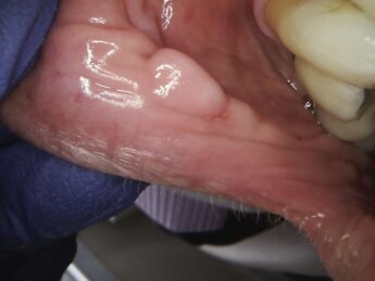
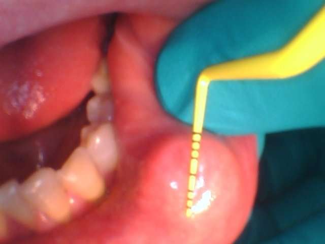
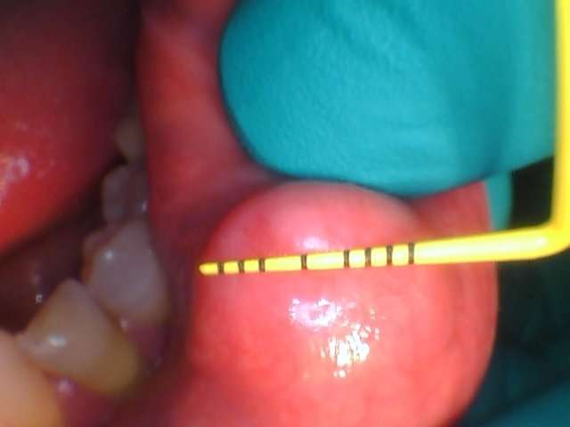
A patient came to Dr. David Stall because she wanted to have a sizable lesion on her lower left lip removed. She had seen an oral surgeon that discussed using a scalpel and making a football-shaped incision that would possibly pull on her lip when sutured. She wanted to avoid this. The patient’s lesion seems firm but was raised, moveable, not painful, and she would often accidentally bite it. It was sized 10 x 10 mm as seen in Figures 1a and 1b. Dr. Stall discussed the use of the laser to release a flap and then the mass underneath.
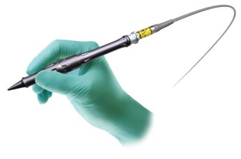
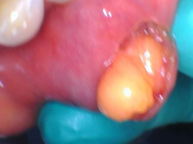
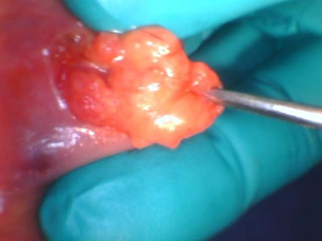
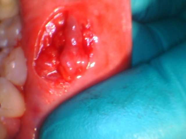
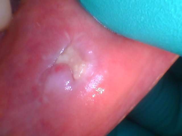
The patient was given local anesthesia and Dr. Stall using his LightScalpel CO2 laser (set at 3 Watts, 20 Hz, 1500 MW, in SuperPulse mode), made a bloodless incision through the lip to access the lesion. The lesion was then excised (See Figures 2-4). He thought it was a lipoma and sent it to the lab for a pathology report. Four sutures (chromic gut 5-0) were placed to minimally approximate the margins of the large lesion without affecting the contours of the outer lip. Due to the hemostatic ability of the CO2 laser, the center of the surgical wound was left to heal by secondary intention.
Compared to a conventional scalpel, the use of the LightScalpel CO2 laser in this lipoma removal offered several benefits. Some of these benefits include cutting in a non-contact mode (to avoid mechanical trauma to the tissue) and without bleeding and minimized postoperative pain, edema, and inflammation.[1]
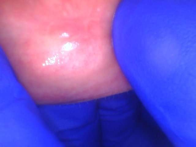
The pathology report confirmed it was a non-cancerous lipoma. Recovery was uneventful and the patient was very happy with the results.
About the Author

References
- Levine R, Vitruk P. “Enhanced hemostasis and improved healing in CO2 laser-assisted soft tissue oral surgeries.” Implant Practice US. 2015;8(3):34-37.